How Advanced Imaging Solutions Are Improving Visualization in Minimally Invasive Surgery
How Advanced Imaging Solutions Are Improving Visualization in Minimally Invasive Surgery

Share:
Share
Visualisation Is at the Heart of Minimally Invasive Surgery
Minimally invasive surgery has grown steadily over the past two decades, replacing many open procedures across general surgery, gynecology, urology, and other specialties. This shift brings a central challenge: surgeons operate through small access points while relying entirely on a monitor to see the surgical field. Unlike open surgery, there is no direct line of sight, no depth cues from natural vision, and no ability to physically feel every structure. This makes the quality of the visual feed a critical factor in how confidently and precisely a surgeon can work.
Research on laparoscopic imaging shows how crucial image quality is, and how good visualization can affect surgical work, especially when you have to spot anatomical structures, or do fine dissection steps. As the procedures get more complicated, imaging solutions are moving far past the usual camera and monitor set up, they now provide sharper and more detailed views across the whole operation. In other words, it’s not only about seeing, it’s about seeing clearly enough to guide the next move.
From Standard HD to 4K: The Evolution of Surgical Imaging
Surgical imaging has progressed through several distinct generations. The early laparoscopic imaging system setups leaned on standard HD, and then shifted to Full HD systems which gave sharper detail with improved color accuracy. More recently, 4K imaging system technology has become common in operating rooms, and ultra high-definition formats keep on emerging.
Each step up in resolution can help show finer anatomical details more clearly, tissue planes, blood vessels, surgical instruments, and those subtle shifts in how tissue looks that might otherwise slip by. There’s also a systematic review that talks about how 4K technology got developed and what role it could play in strengthening visual cues like shadows, color, contrast, and depth perception. That said, going higher resolution by itself doesn’t always mean the outcome will be better for every single case. What it does is expand the visual information available to the surgeon, the actual clinical benefit still depends on the procedure being performed, how the system is configured, and the surgeon's own experience with the technology.
Better Visualisation Means Better Understanding of the Surgical Field
Clearer imaging has real practical value in the operating room. It supports better recognition of anatomical landmarks, sharper differentiation between similar-looking structures, and improved visibility during dissection, all of which can build a surgeon's confidence when navigating complex or delicate anatomical regions. Colour reproduction and contrast play just as important a role here as raw resolution. Research in colorectal laparoscopic surgery has linked improved visualization with clearer anatomical landmarks and potentially more precise dissection. It's important to understand that surgical visualization isn't simply a matter of "more pixels." The overall quality of what a surgeon sees depends on the camera, the optics, the light source, the image processor, the display, and how well all these components are integrated into a single imaging chain.
2D, 3D, and 4K: Choosing the Right Visualisation Approach
There's no single imaging technology that works best for every procedure or every surgeon. Options today generally fall into a few categories:
2D HD – an established, widely used standard
2D 4K – higher resolution with more detailed visualization
3D HD – adds stereoscopic depth perception
Advanced imaging – combines high resolution with additional image-enhancement technologies
Studies comparing 3D and 4K systems have produced mixed findings, with results varying depending on the task and the outcome being measured. In practice, the right choice often comes down to clinical requirements, surgical workflow, individual surgeon preference, and the infrastructure already available in the operating room.
The Role of Advanced Camera and Image Processing Technologies
Modern imaging solutions extend well beyond camera resolution alone. High-resolution surgical cameras are paired with advanced image processing to deliver improved colour reproduction, better contrast enhancement, and consistent image optimization across the length of a procedure. Stable, reliable video output, along with smooth integration with surgical displays and recording systems, matters just as much as the initial image capture. This is where a carefully engineered endoscopic imaging system shows its value as a complete solution rather than something that is a single standalone device. For hospitals evaluating new equipment, this points to a broader consideration: selecting an imaging system that fits naturally into the operating room's existing workflow, not just one with impressive specifications on paper.
What Hospitals Should Consider When Choosing an Imaging Solution
Selecting the right system involves more than comparing resolution numbers. Hospitals should consider:
Image resolution and camera quality
Colour reproduction and image processing capabilities
Light source compatibility
Display quality
Ease of integration with existing systems
Recording and documentation capabilities
Upgradeability
Service and technical support
Compatibility with existing OT infrastructure
Matrix Medicals: Supporting Modern Surgical Visualisation
Matrix Medicals provides medical technology solutions, designed for the modern operating rooms. Their portfolio covers endoscopic imaging system offerings, and a wider set of OT technologies like the Lucerna Endoscopic Camera System. As minimally invasive surgery keeps changing and moving forward, Matrix Medicals tries to back hospitals with imaging and operating room tech that fits the needs of today’s surgical workflows, and it connects in a natural way with their bigger OT ecosystem of OT lights, OT tables, electrosurgical units, and OR integration solutions.
Conclusion: The Future of Minimally Invasive Surgery Is Clearer Visualization
Visualization remains fundamental to the practice of minimally invasive surgery. Imaging technology has progressed from conventional HD toward 4K, 3D, and increasingly sophisticated image-enhancement capabilities, and this evolution shows no signs of slowing. The future will likely bring even greater integration of imaging, data, AI, and OR systems working together. For hospitals, the key takeaway is to evaluate imaging solutions based on the complete surgical workflow, not resolution figures alone.
Looking to explore advanced endoscopic and laparoscopic imaging solutions for your operating room? Connect with Matrix Medicals to discuss the right imaging approach for your surgical environment.
Visualisation Is at the Heart of Minimally Invasive Surgery
Minimally invasive surgery has grown steadily over the past two decades, replacing many open procedures across general surgery, gynecology, urology, and other specialties. This shift brings a central challenge: surgeons operate through small access points while relying entirely on a monitor to see the surgical field. Unlike open surgery, there is no direct line of sight, no depth cues from natural vision, and no ability to physically feel every structure. This makes the quality of the visual feed a critical factor in how confidently and precisely a surgeon can work.
Research on laparoscopic imaging shows how crucial image quality is, and how good visualization can affect surgical work, especially when you have to spot anatomical structures, or do fine dissection steps. As the procedures get more complicated, imaging solutions are moving far past the usual camera and monitor set up, they now provide sharper and more detailed views across the whole operation. In other words, it’s not only about seeing, it’s about seeing clearly enough to guide the next move.
From Standard HD to 4K: The Evolution of Surgical Imaging
Surgical imaging has progressed through several distinct generations. The early laparoscopic imaging system setups leaned on standard HD, and then shifted to Full HD systems which gave sharper detail with improved color accuracy. More recently, 4K imaging system technology has become common in operating rooms, and ultra high-definition formats keep on emerging.
Each step up in resolution can help show finer anatomical details more clearly, tissue planes, blood vessels, surgical instruments, and those subtle shifts in how tissue looks that might otherwise slip by. There’s also a systematic review that talks about how 4K technology got developed and what role it could play in strengthening visual cues like shadows, color, contrast, and depth perception. That said, going higher resolution by itself doesn’t always mean the outcome will be better for every single case. What it does is expand the visual information available to the surgeon, the actual clinical benefit still depends on the procedure being performed, how the system is configured, and the surgeon's own experience with the technology.
Better Visualisation Means Better Understanding of the Surgical Field
Clearer imaging has real practical value in the operating room. It supports better recognition of anatomical landmarks, sharper differentiation between similar-looking structures, and improved visibility during dissection, all of which can build a surgeon's confidence when navigating complex or delicate anatomical regions. Colour reproduction and contrast play just as important a role here as raw resolution. Research in colorectal laparoscopic surgery has linked improved visualization with clearer anatomical landmarks and potentially more precise dissection. It's important to understand that surgical visualization isn't simply a matter of "more pixels." The overall quality of what a surgeon sees depends on the camera, the optics, the light source, the image processor, the display, and how well all these components are integrated into a single imaging chain.
2D, 3D, and 4K: Choosing the Right Visualisation Approach
There's no single imaging technology that works best for every procedure or every surgeon. Options today generally fall into a few categories:
2D HD – an established, widely used standard
2D 4K – higher resolution with more detailed visualization
3D HD – adds stereoscopic depth perception
Advanced imaging – combines high resolution with additional image-enhancement technologies
Studies comparing 3D and 4K systems have produced mixed findings, with results varying depending on the task and the outcome being measured. In practice, the right choice often comes down to clinical requirements, surgical workflow, individual surgeon preference, and the infrastructure already available in the operating room.
The Role of Advanced Camera and Image Processing Technologies
Modern imaging solutions extend well beyond camera resolution alone. High-resolution surgical cameras are paired with advanced image processing to deliver improved colour reproduction, better contrast enhancement, and consistent image optimization across the length of a procedure. Stable, reliable video output, along with smooth integration with surgical displays and recording systems, matters just as much as the initial image capture. This is where a carefully engineered endoscopic imaging system shows its value as a complete solution rather than something that is a single standalone device. For hospitals evaluating new equipment, this points to a broader consideration: selecting an imaging system that fits naturally into the operating room's existing workflow, not just one with impressive specifications on paper.
What Hospitals Should Consider When Choosing an Imaging Solution
Selecting the right system involves more than comparing resolution numbers. Hospitals should consider:
Image resolution and camera quality
Colour reproduction and image processing capabilities
Light source compatibility
Display quality
Ease of integration with existing systems
Recording and documentation capabilities
Upgradeability
Service and technical support
Compatibility with existing OT infrastructure
Matrix Medicals: Supporting Modern Surgical Visualisation
Matrix Medicals provides medical technology solutions, designed for the modern operating rooms. Their portfolio covers endoscopic imaging system offerings, and a wider set of OT technologies like the Lucerna Endoscopic Camera System. As minimally invasive surgery keeps changing and moving forward, Matrix Medicals tries to back hospitals with imaging and operating room tech that fits the needs of today’s surgical workflows, and it connects in a natural way with their bigger OT ecosystem of OT lights, OT tables, electrosurgical units, and OR integration solutions.
Conclusion: The Future of Minimally Invasive Surgery Is Clearer Visualization
Visualization remains fundamental to the practice of minimally invasive surgery. Imaging technology has progressed from conventional HD toward 4K, 3D, and increasingly sophisticated image-enhancement capabilities, and this evolution shows no signs of slowing. The future will likely bring even greater integration of imaging, data, AI, and OR systems working together. For hospitals, the key takeaway is to evaluate imaging solutions based on the complete surgical workflow, not resolution figures alone.
Looking to explore advanced endoscopic and laparoscopic imaging solutions for your operating room? Connect with Matrix Medicals to discuss the right imaging approach for your surgical environment.
Share:
Share
RELATED ARTICLES
Read more from our blogs

Why Is Zero Positioning Important in Modern Operation Theatre Tables?
Why Is Zero Positioning Important in Modern Operation Theatre Tables?

Why Is Saline-Compatible Cautery Machine Important in Urological Surgery?
Why Is Saline-Compatible Cautery Machine Important in Urological Surgery?

How Medical Laboratory Technology Is Improving the Speed and Accuracy of Diagnostics
How Medical Laboratory Technology Is Improving the Speed and Accuracy of Diagnostics
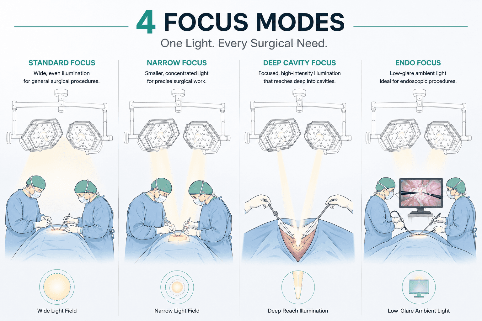
What Are the Different Focus Modes in Modern Surgical Theatre Lights?
What Are the Different Focus Modes in Modern Surgical Theatre Lights?

Products
Get in touch


info@matrixmedicals.in

+91 7304411451
+91 9821235552
Copyright © 2026 Matrix Medicals Pvt. Ltd., All rights reserved.
Products
Get in touch
info@matrixmedicals.in
+91 7304411451
+91 9821235552
Copyright © 2026 Matrix Medicals Pvt. Ltd., All rights reserved.
Contact Us
Contact Us
Contact Us